Effects of exercise type on estrogen, tumor markers, immune function, antioxidant function, and physical fitness in postmenopausal obese women
Article information

Abstract
This study aims to identify the effects of exercise type on estrogen, tumor markers, immune function, antioxidant function, and physical fitness in postmenopausal obese women. The subjects were 30 post-menopausal obese women with body fat percentage higher than 30%. Participants were divided into aerobic exercise group (n=10; age, 53.70±3.37 years), resistance exercise group (n=10; age, 52.20±2.15 years), and control group (n=10; age, 52.50±2.68 years). Estrogen and growth hormone showed no significant difference in the aerobic exercise group, resistance exercise group, and control group. Tumor marker alpha-fetoprotein was increased in the aerobic exercise, resistance exercise, and control groups (P<0.01). The metabolic syndrome risk factor was decreased in the aerobic and resistance exercise groups, which was shown by the reduction of weight (P<0.001), body fat percentage (P<0.001), waist circumference (P<0.05), and increase of high density lipoprotein-cholesterol (P<0.001). natural killer cell activity was increased in the aerobic exercise group, resistance exercise group, and control group (P<0.001). Oxidative stress was decreased in the aerobic exercise group, resistance exercise group, and control group (P<0.001). Maximum oxygen uptake was increased in the aerobic and resistance exercise groups, but aerobic exercise was more effective (P<0.05). Knee isokinetic extensor muscle was increased in both the aerobic and resistance exercise groups (P<0.001). Aerobic and resistance exercise of postmenopausal obese women can be considered an effective intervention program to prevent metabolic syndrome and improve physical fitness.
INTRODUCTION
Menopause is a type of aging process during which the secretion of the female hormone estrogen is decreased due to a decline of follicular function, and the quality of life and physical functions deteriorate due to menopausal symptoms (Nelson, 2008). In particular, postmenopausal women are known to have a higher chance of having cancer than premenopausal women (Rosano et al., 2007). With recent emphasis on the importance of early discovery of cancer, researches on tumor markers have been actively done. Tumor markers are used to selecting and tracking the early diagnosis of cancer and to predict the pre- and postoperative prognosis, but due to low sensitivity and specificity for tumor diagnosis, they appear to be increased or get detected even when one does not have cancer (Perkins et al., 2003). In other words, most cancers are easy to track down but tumor markers for liver cancer and breast cancer, which often start around menopausal stage, have been reported to be diagnostically useful (Sturgeon, 2002).
Also, postmenopausal women have reduced production of estrogen, which serves a cardioprotective function; this event leads to a higher risk of developing cardiovascular disease (Knopp, 1988). In other words, estrogen deficiency due to menopause accelerates the accumulation of visceral fat, which increases the risk of metabolic syndrome, in which abdominal obesity and insulin resistance play important roles (Carr, 2003). Moreover, estrogen plays an important role in lymphocyte activation, but menopause decreases the efficiency of cell-mediated and antibody-mediated responses by thymic atrophy (Pernis, 2007; Ryan et al., 2005).
On the other hand, the pathogenesis of cancer, cardiovascular disease, and impaired immune function can be attributed to the deterioration of cell damage and antioxidant activity by free radicals (Valko et al., 2007) and is significantly affected by physical inactivity (Hamilton et al., 2007). However, aerobic exercises suggested for menopause stage are known to be effective in the prevention of cancer and metabolic syndrome, improvement of immune system and antioxidant activity (Bruunsgaard and Pedersen, 2000; Friedenreich, 2001; Hartmann and Niess, 1988; Oliveria and Christos, 1997). In addition, since the skeletal muscles are decreased during menopausal period, resistance exercise is also important. Resistance exercise has been shown to be effective in improving myocardial function and preventing chronic diseases (Bemben et al., 2000; Hurley et al., 1988).
However, studies that have compared and analyzed changes in estrogen, tumor marker, metabolic syndrome indicator, immune function, antioxidant function and physical fitness between aerobics and exercises among obese postmenopausal women are highly limited. Therefore, the aim of this study is to compare and analyze the effect of 12-week-long aerobic and resistance exercises on estrogen, tumor marker, metabolic syndrome indicator, immune function, antioxidant function and physical fitness in postmenopausal obese women.
MATERIALS AND METHODS
Subjects
For the subjects of this study, 30 obese postmenopausal women with a body fat percentage of 30% or higher were registered at Seoul regional sports center, and they were randomly distributed into aerobic exercise group (n=10), resistance exercise group (n=10), and control group (n=10). Physical characteristics of the subjects are shown in Table 1. The exclusion criteria were those who were taking medication due to heart disease, those who have been taken sex hormonal drugs, those under estrogen replacement therapy, and those who have experienced chest pain, dyspnea, and arrhythmia during exercise. This study was approved by the Institutional Review Board of the Korea National Sport University’s Bioethics Committee (approval number: 1263-201706-BR-005-04).
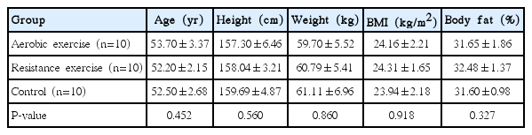
Characteristics of subjects
Measurement items and methods
Height and weight were measured using an automatic body meter (Jenix, Seoul, Korea). The body mass index (BMI) was calculated by dividing the measured body weight (kg) by the square (m2) of the height (cm). The waist circumference was measured up to 0.1 cm in the upright posture from the lower end of the rib and the midpoint of the upper iliac crests. The body fat percentage was measured using a body fat analyzer (X-Scan II, Jawon Medical, Gyeongsan, Korea) following the bioelectrical impedance method.
The blood pressure measurement was done by measuring systolic and diastolic blood pressures in the left brachial artery at the same height as the heart by using a mercury blood pressure meter after at least 10 min of resting.
Maximum oxygen uptake (VO2max) was measured to show cardiovascular strength by performing an exercise load test based on a modified version of Balke protocol using an automatic breathing gas analysis system (Q4500, Quinton, Bothell, WA, USA). The criteria for ending the exercise load test were when the oxygen consumption did not increase even when the exercise load was increased, when the exercise self-conscious level was 17 or higher, when the heart rate was ±5 than the age-dependent predicted maximum heart rate by age, and when the respiratory exchange rate was higher than 1.15.
To measure the maximal strength, relative strength, and extensor motion range of the knee extensions and flexor muscles, the test was performed using isokinetic equipment (System 3 Pro, Biodex, Shirley, NY, USA). The measurement was done 3 times at the left and right at the speed of 60°/sec after matching the subject’s knee motion axis and dynamometer axis and by fixating the trunk and thigh with Velcro to minimize the motion of other body parts.
Blood samples were collected from the vein after confirming the 10-hr fasting state. The collected blood was centrifuged at 3,000 rpm for 10 min and stored at −70°C. Blood analysis items and methods are as follows. Estrogen was analyzed via radioimmunoassay. Growth hormone was analyzed using chemiluminescence immunoassay. Tumor markers alpha-fetoprotein (AFP), carcinoembryonic antigen (CEA), cancer antigen 125 (CA 125), and CA 153 were analyzed by radioimmunoassay. The triglyceride, high-density lipoprotein cholesterol (HDL-C), and fasting state blood sugar were analyzed by enzymatic method, while low density lipoprotein cholesterol (LDL-C) was done by homogeneous enzymatic colorimetric assay. To assess the extent of insulin resistance, the formula of Matthews et al. (1985) was used: “HOMA-IR=[fasting plasm insulin (μU/mL)×fasting plasm glucose (mg/dL)]/405.” Natural killer (NK) cells were analyzed by enzyme-linked immunosorbent assay, whereas immuneglobulins (IgA, IgG, and IgM) were analyzed by turbidometry method. Total antioxidant status (TAS) and total oxidant status (TOS) were analyzed using a colorimetry method.
Exercise program
The aerobic and resistance exercise program of this study was conducted 3 times a week for 12 weeks after the subjects were given an education on exercise method and intensity setting, as well as an adjustment period. The heart rate reserve (HRR) was set using the Karvonen et al. (1957) formula for the aerobic exercise intensity. Resistance exercise intensity was set using the indirect measurement method of one repetition maximum or 1 RM (Fleck and Kramer, 1987). Exercise time included 10 min of pre-exercise, 40 min of main exercise, and 10 min of finishing exercise. The pre-exercise consisted of 5 min of walking and 5 min of stretching, while the finishing exercise included 10 min of stretching. The main exercise was as follows.
The aerobic exercise started with walking on a treadmill at different speeds and slopes at the HRR of 60%–80%. During the exercise, the subject wore a heart rate monitor (Polar, Kempele, Finland) to check whether the exercise intensity has been reached. Resistance exercise included bench press, lat-pull down, triceps push-down, dumbbell curl, sit-up, squat, leg extension, leg flexion, and leg press at 1RM of 60%. The exercises were repeated 8–12 times for 3 sets. The resting time between two exercise types was 1-min long.
Data analyses
The mean and standard deviation per measured item from this study’s data were calculated using the IBM SPSS ver. 18.0 (IBM Co., Armonk, NY, USA). Two-way analysis of variance with repeated measures was performed to verify the mean difference. The significance level (α) of this study was set to P<0.05.
RESULTS
Change in estrogen and growth hormone
Estrogen and growth hormone change due to aerobic exercise and resistance exercise is shown in Table 2. Value of estrogen and growth hormone did not have a significant difference between groups and measuring time points and there was no interaction effect.

Change of estrogen and growth hormone
Change in tumor markers
Tumor markers change due to aerobic exercise and resistance exercise is shown in Table 3. The value of AFP tended to increase in each of aerobic exercise group, resistance exercise group, and control group, while significant difference was shown between measuring time points (P<0.05). There were no interaction effects. On the other hand, for CEA, CA 125, and CA 153, values did not have significant difference between groups and measuring time points. There were no interaction effects.
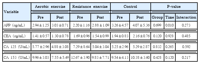
Change of tumor markers
Change in metabolic syndrome risk factors
Change in metabolic syndrome risk factors due to aerobic exercise and resistance exercise is shown in Table 4. The value of weight tended to decrease in the aerobic exercise group and resistance exercise group, and there was significant difference between measuring time points (P<0.001). There was also interaction effect (P<0.001). The value of BMI tended to decrease in the aerobic exercise group and resistance exercise group, and there was significant difference between measuring time points (P<0.01). There was also interaction effect (P<0.01). The value of body fat tended to decrease in the aerobic exercise group and resistance exercise group, and there was significant difference between measuring time points (P<0.05). There was no interaction effect. The value of waist circumference tended to decrease in the aerobic exercise group and resistance exercise group, and there was significant difference between measuring time points (P<0.05). There was no interaction effect.
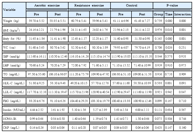
Change of metabolic syndrome risk factors
The value of systolic blood pressure and diastolic blood pressure did not have a significant difference between measuring time points. There was no interaction effect. The value of triglyceride did not have significant difference between groups and measuring time points. There was no interaction effect. The value of HDL-C tended to increase in the aerobic exercise group and resistance exercise group, and there was significant difference between measuring time points (P<0.01). There was no interaction effect. The value of LDL-C did not have a significant difference between groups and measuring time points. There were no interaction effects.
The value of fasting blood glucose, insulin, homeostasis model assessment of insulin resistance index (HOMA-IR), and C-reactive protein (CRP) did not have significant difference between groups and measuring time points. There were no interaction effects.
Change in immune function
Change in immune function due to aerobic exercise and resistance exercise is shown in Table 5. The value of NK cell activity tended to increase in all aerobic exercise groups, in the resistance exercise group, and in the control group. There was significant difference between measuring time points (P<0.001). There was no interaction effect. The value of IgA did not have a significant difference between measuring time points. There was no interaction effect. The value of IgG tended to decrease in the aerobic exercise group and resistance exercise group but increase in the control group. There was significant difference between measuring time points (P<0.05), and also interaction effect (P<0.05). Value of IgM tended to decrease in the aerobic exercise group and resistance exercise group. There was significant difference between measuring time points (P<0.001). There was no interaction effect.

Change of immune function
Change in antioxidant function
Change in antioxidant function due to aerobic exercise and resistance exercise is shown in Table 6. TAS, which is a measurement indicator for antioxidant capacity, did not show significant difference in value between groups and measuring time points. There was no interaction effect. The value of TOS, which is a measurement indicator for oxidative stress, tended to decrease in the aerobic exercise group, resistance group, and control group. There was significant difference between measuring timing points (P<0.001). There was no interaction effect.

Change of antioxidant function
Change in physical fitness
Change in cardiorespiratory fitness and isokinetic function of knee joint due to aerobic exercise and resistance exercise is shown in Table 7. The value of VO2max increased in the aerobic exercise group and decreased in the control group. There was no significant difference between measuring time points. There was interaction effect (P<0.05).

Change of cardiorespiratory fitness and knee isokinetic function
The value of left knee joint isokinetic extensor tended to increase in the aerobic exercise group and resistance exercise group. There was significant difference between measuring time points (P<0.001). There was interaction effect (P<0.05). The value of right knee joint isokinetic extensor tended to increase in the aerobic exercise group and resistance exercise group. There was significant difference between measuring time points (P<0.001). There was interaction effect (P<0.05). The value of left and right knee joint isokinetic flexors did not have a significant difference between groups and measuring time points, and there was no interaction effect.
DISCUSSION
This study assessed the effects of exercise type on estrogen, tumor markers, immune function, antioxidant function and physical fitness in postmenopausal obese women. As a result, aerobic and resistance exercises did not affect changes in estrogen and growth hormone. The tumor marker AFP was increased, while IgG and IgM were decreased. On the other hand, the exercises appeared to be effective in the reduction of obesity indicators of metabolic syndrome, increase of HDL-C, NK cell activation, reduction of oxidative stress, and improvement of physical fitness. However, no variables except for VO2max showed significant changes between the aerobic and resistance exercises. Moreover, the control group of this study showed an increase of AFP and NK cell, as well as reduction of oxidative stress.
Menopause comes with various physiological changes due to the cease of menstruation from ovarian functional failure. During the menopausal state, the decrease of estrogen secretion is one representative symptom. On the other hand, if postmenopausal women are exposed to high estrogen level, it can induce breast cancer (Cauley et al., 1989). This study revealed that aerobic, resistance, and control exercise groups all showed an increase in estrogen level but with no significant differences. Similar to this study, Robles et al. (2012) also tested premenopausal and postmenopausal women by having them do aerobic exercises for 6 months but no changes were found in their estrogen level. According to Smith et al. (2013), a 16-week aerobic exercise did not affect the change in estrogen level in postmenopausal women, but it was effective in the prevention of breast cancer by inducing the increase of estrogen metabolites 2-hydroxyestrone and reduction of 16α-hydroxyestrone. Therefore, the study on the exercise type and estrogen metabolism in postmenopausal women needs to be studied in further detail. In this study, growth hormone was decreased in aerobic exercise and control group but increased in aerobic exercise group, and no significant differences were observed. Both aerobic and resistance exercises showed to have similar effects on the stimulation of growth hormone secretion, but since the amount of exercise decided by the intensity and duration is a critical factor, future studies should be done by taking this into consideration (Wideman et al., 2002).
Obesity after menopause increases the risk of having cancer (Song et al., 2008). Tumor markers are useful diagnostic tools that can be used for the early detection of cancer, the determination of treatment efficacy, and the possibility of recurrence (Perkins et al., 2003). According to cohort studies on cancer development, exercise is protective against cancer (Friedenreich, 2001). The results from this study show that AFP, a tumor marker of liver cancer, was significantly increased in aerobic exercise, resistance exercise, and control group. On the other hand, CEA, cervical CA 125, and breast CA 153 were decreased but not significantly changed. Similar to this study, 8 weeks of aerobic exercise was shown to be ineffective in inducing changes in the CEA and CA 153 among breast cancer patients (Esfahbodi et al., 2018). However, tumor markers have a low specificity and, thus, play limited role in cancer screening. Furthermore, various physiological factors are known to affect tumor marker levels. In particular, the increase of AFP observed in this study may be elevated due to various liver diseases such as hepatitis, liver cirrhosis, and liver tumors (Christiansen et al., 2001). Therefore, future studies are needed to assess the exercise and tumor marker in obese menopausal women from multifaceted points of view.
The low prevalence of cardiovascular disease in premenopausal women is due to the protective effects of estrogen against cardiovascular disease (Gavin et al., 2009). However, menopausal estrogen deprivation results in abdominal fat accumulation and insulin resistance, which are major risk factors for the development of metabolic syndrome (Lobo, 2008). The results of this study showed that aerobic exercise and resistance exercise were effective in decreasing obesity indicators (weight, BMI, waist circumference, body fat percentage) and increasing HDL-C among metabolic syndrome risk factors. However, no significant changes were observed in blood pressure, triglyceride, insulin resistance level, and inflammatory marker CRP. These results are partially in line with studies that have shown that aerobic exercise in postmenopausal women fights against metabolic syndrome risk factors while reducing HOMA-IR (Earnest et al, 2013; Kim and Kim, 2012) and that metabolic syndrome z-score is decreased by resistance exercise (Conceição et al., 2013). In general, postmenopausal women have increased body fat and abdominal obesity due to the imbalance between nutritional intake and consumption, and a decrease in estrogen secretion promotes this process (Poehlman, 2005). In this study, aerobic and resistance exercise reduced weight and waist circumference without significant changes in the level of estrogen, which causes fat accumulation in postmenopausal obese women. This is due to that fact that calorie consumption-inducing exercise decomposes the triglyceride stored in adipocytes into free fatty acids which is to be used as energy source by muscles.
Exercise is effective in preventing cancer and infectious diseases by improving cell-mediated immune responses and adaptive immune responses (Karacabey et al., 2005). This study showed that NK cell activity was significantly increased in aerobic exercise, resistant exercise, and control group. On the other hand, IgA was not changed, but IgG and IgM were significantly decreased in aerobic exercise group and resistant exercise group. Campbell et al. (2008) reported that aerobic exercise increased NK cell, IgA, and IgG levels among postmenopausal women, but the changes were not significant. The increased NK cell activity shown in this study suggests that it may be effective in preventing cancer among postmenopausal women. However, the reason that NK cell activity was increased in both exercise and control groups may be due to external factors such as nutritional intake, stress level, and sleep status. In addition, IgG and IgM, which are known to be increased by exercise, were decreased. This may be due to immunoglobulin inhibition occurring through an increase in male hormone testosterone in the menopausal state (Giltay et al., 2000). Moreover, immunoglobulin is affected by exercise intensity, stress level, and nutritional status (Pedersen and Hoffman, 2000). Therefore, it is necessary to analyze the exercise and immunoglobulin promoting effect in postmenopausal obese women by considering these variables in the future.
Development of cancer, cardiovascular disease, and deterioration of immune function in postmenopausal women is suggested to be due to increased free radical production and decreased antioxidant function. Oxidative stress caused by free radicals results to oxidative damage of cellular proteins, lipids, and nucleic acids (Andriollo-Sanchez et al., 2005). This study showed that the TAS was increased in the aerobic exercise group, resistant exercise group, and control group, but there was no significant difference. It was found to be effective in reducing TOS, which is an oxidative stress indicator. In a previous study, it was reported that aerobic exercise increases antioxidant enzyme activity while decreasing oxidative stress (Attipoe et al., 2008; Jarrete et al., 2014). Similarly, aerobic and resistance exercise among postmenopausal women can be beneficial in reducing oxidative stress, which is used as a disease pathogenesis measure. However, in this study, the increase of antioxidant activity and increase of oxidative stress level were also observed in the control group. This may be due to the fact that the antioxidative role of various nutritional factors like vitamin C, vitamin E, albumin, and beta-carotene was not appropriately considered in this study. It seems necessary to identify the effect of exercise by considering this factor in the future.
On the other hand, low cardiorespiratory fitness and decreased muscle strength are risk factors for cardiovascular disease (García-Hermoso et al., 2018; Wessel et al., 2004). A number of studies reported that aerobic and resistance exercise are effective in increasing cardiorespiratory fitness and muscle strength among postmenopausal women (Karacan, 2010; Oliveira et al., 2015; Orsatti et al., 2008). This study showed that VO2max was increased in all exercise groups but more significantly in the aerobic exercise group. The isokinetic knee extensor muscles were increased in both the aerobic exercise group and resistance exercise group. Recently, Robinson et al. (2017) have shown that aerobic interval training exercise is effective in increasing VO2max and muscle strength, while resistance exercise does not affect VO2max enhancement. The difference in the cardiovascular fitness and muscular strength enhancement between aerobic and resistance exercises were also observed in this study, but it can suggest it as an effective method for increasing physical fitness, which is important for the prevention of cardiovascular disease in postmenopausal obese women.
The limitation of this study is that it is difficult to generalize the data because subjects were selected in postmenopausal obese women from one single region. Moreover, this study did not include the amount of physical activity, smoking, drinking, dietary habits, and stress level, indicating that the study did not take the effect of these factors on the measuring variables into consideration.
In conclusion, aerobic exercise and resistance exercise are not effective in inducing changes in the estrogen secretion in postmenopausal obese women but may be considered as a basis for intervention for the prevention of metabolic syndrome and improvement of physical fitness. In order to verify the effect of each variable, future studies are needed to be carried out by taking intensity, amount, and duration of aerobic and resistance exercise.
Notes
CONFLICT OF INTEREST
No potential conflict of interest relevant to this article was reported.
ACKNOWLEDGMENTS
This work was supported by the National Research Foundation of Korea (NRF) grantfunded by the Korea government (2017S1 A5A2A01026057).