INTRODUCTION
Peripheral nerves are often damaged by crush, compression, stretching, avulsion, and division. The injury of nerve causes loss of function of internal organs and muscles. Following sciatic crushed nerve injury (SCI), pain-related gait and swelling are displayed in the affected limb.
Ascending central pathways transmit nociceptive information to the pain-related brain areas (Price, 2000). Of these, periaqueductal gray (PAG) and paraventricular nucleus (PVN) are important brain sites involved in ascending pain transmission. It receives afferent from nociceptive neurons in the spinal cord and send projection to the thalamic nuclei that process nociception.
Immediate early gene c-fos has been considered as a marker for stimuli-induced metabolic change of neurons. Expression of c-Fos is enhanced by various stimuli and c-Fos is a sensitive marker for the neural activation (de Medeiros et al., 2003; Narita et al., 2003). c-Fos expression in the PAG and PVN suggests that neuronal activation occurs in a location-specific manner (Belevych et al., 2010).
Neurofilament (NF) is an intermediate filament that found in neurons. NF is a major component of the neuronal cytoskeleton, and provides structural support for the axon (Park et al., 2013). NF-200 is one of the types of cytoskeletal protein and NF-200 plays a role in the stabilization and maturation of pre-existing connections (Kriz et al., 2000).
Brain-derived neurotrophic factor (BDNF) is a member of the neurotrophic family and BDNF promotes the survival of neurons during development. BDNF modulates many morphological changes such as dendritic arborization, remodeling of axon and dendrite, and synaptogenesis (Sallert et al., 2009; Yacoubian and Lo, 2000). BDNF up-regulation represents neuronal regeneration and improvement of locomotor function (Jung et al., 2016; Kim et al., 2017).
The scientific functional index (SFI) is a mathematical formula to represent parameters of normal and experimental footprints. SFI provides information of gait function associated with sensory-motor connections (de Medinaceli et al., 1982). SFI has been revised several times to become a simpler and more reliable index. SFI is used to assess functional improvement after SCI (Byun et al., 2005; Lee et al., 2018).
Acupuncture has been used for to treat various maladies, especially pain (Cherkin et al., 2003). Electroacupuncture (EA) is a modified acupuncture technique, as its name implies, that utilizes electrical stimulation. Analgesic effect of EA on different types of acute pain and persistent inflammatory pain has been documented in rodents and humans (Baek et al., 2005; Chang et al., 2004; Gim et al., 2011). Zusanli acupoint (ST36) is the most commonly used acupoint for the pain control and immune regulation (Chang et al., 2004). EA relieves the behavioral signs of hyperalgesia and allodynia associated with neuropathic pain (Dong et al., 2006; Huang et al., 2004; Gim et al., 2011).
In the present study, we investigated the effect of low-frequency EA on functional recovery following SCI in rats. For this study, immunohistochemistry for c-Fos in the ventral lateral PAG (vlPAG) and PVN and western blot for NF-200 and BDNF in the sciatic nerve were conducted.
MATERIALS AND METHODS
Animals and treatments
Male Sprague-Dawley rats weighing 220±10 g (8 weeks old) were used and the experiment was performed in accordance with the animal care guidelines of the National Institute of Health and the Korean Academy of Medical Sciences. The rats were randomly divided into five groups (n=10 in each group): the sham-operation group, the SCI-induced group, the SCI-induced and nonacupoint acupuncture group, the SCI-induced and ST36-acupoint acupuncture group, the SCI-induced and EA group.
The rats in the acupuncture groups received acupuncture bilaterally at respective site, once a day for 14 days. For acupunctural stimulation, after insertion of stainless acupuncture needles (0.3 mm), the needles were manually rotated clockwise and counter-clockwise 30 times and then kept in place for 10 min. Nonacupoint is located at the side of the hip. ST36-acupoint (Zusanli) is located at the four fingers below the tibialis anterior muscle. The rats in the EA group received 100-Hz electrical stimulation for 10 min once a day during 14 days.
Crush injury on sciatic nerve
To induce crush injury on the sciatic nerve, the previously described surgical procedure was performed (Byun et al., 2005; Lee et al., 2018). Right sciatic nerve was exposed by incision on the gluteal muscle under anesthesia using Zoletil 50 (50 mg/kg, intraperitoneally; Vibac Laboratories, Carros, France). The sciatic nerve was carefully exposed and crushed for 30 sec using a surgical clip (pressure: 125 g; Fine Science Tool Inc., San Francisco, CA, USA). The crushed location was between the sciatic notch and the point of trifurcation.
Walking tract analysis
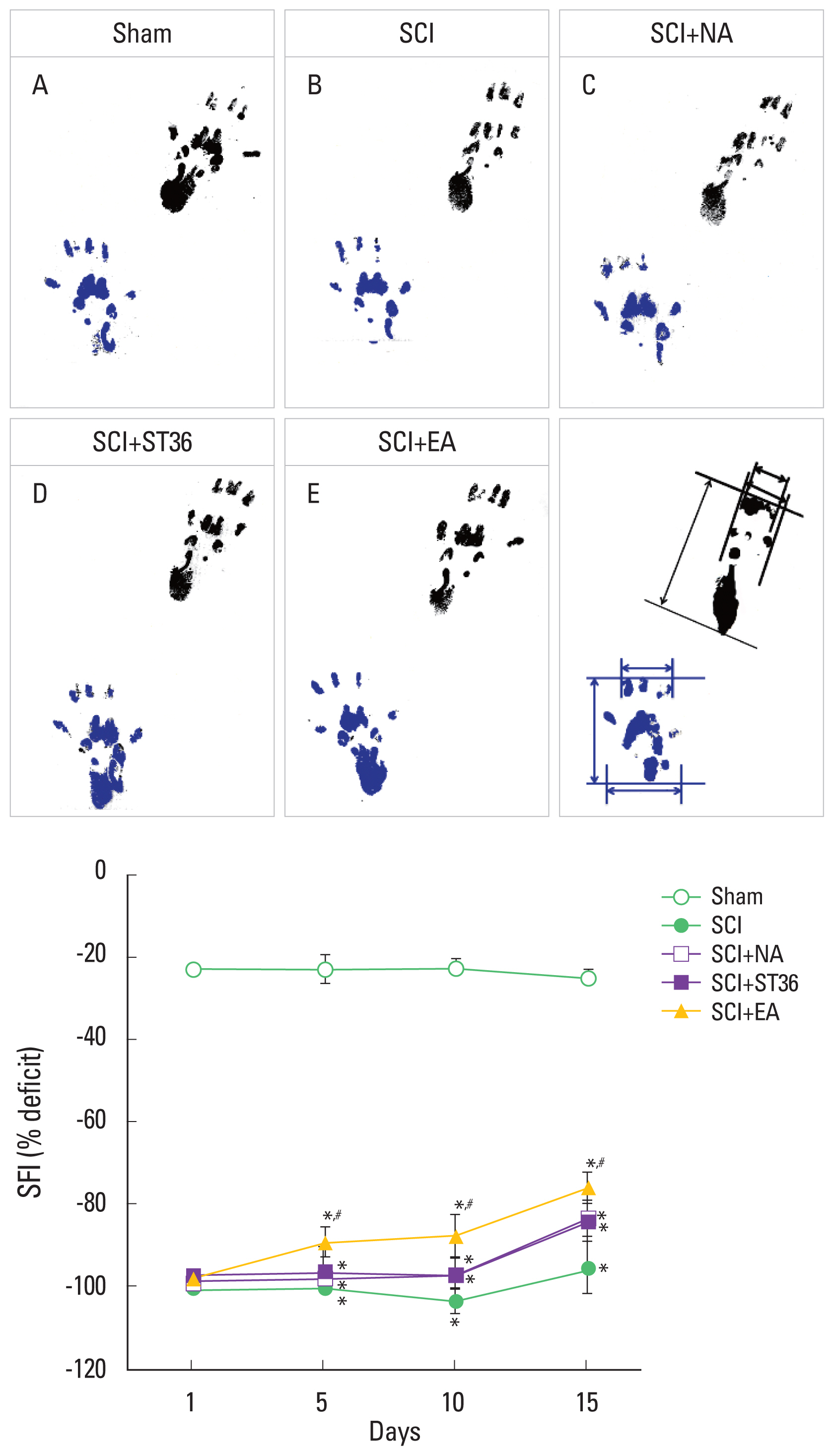
Functional recovery after SCI was quantified by SFI. Examination of the walking patterns was performed 3 times with 1 day intervals through the course of the experiment as the previously described method (Byun et al., 2005; Lee et al., 2018). Foot-prints were recorded in a wooden walking alley (8.2 cm×42 cm) with a darkened goal box at the end. The floor of the alley was covered with white paper. The anatomical landmarks on the hind feet of the rats were smeared with finger point. The rat was allowed to walk down the tract, leaving its foot-prints on the paper.
Following parameter were calculated from the footprints: distance from the heel to the top of the 3rd toe (print length, PL), distance between the 1st to the 5th toe (toe spread, TS), and distance from the 2nd to the 4th toe (intermediary toe spread, IT). These parameters were taken both from the intact left (non-operated) foot (NPL, NTS, and NIT) and from the injured right (experimental) foot (EPL, ETS, and EIT). SFI values were obtained using following equation: SFI=(−38.3±PLF)+(109.5±TSF)+ (13.3±ITF) −8.8. Print length factor (PLF)=(EPL−NPL)/NPL; toe spread factor (TSF)=(ETS−NTS)/NTS; intermediary toe spread factor (ITF)=(EIT−NIT)/NIT. Interpolating identical values of PL, TS, and IT from the right and left hind feet are close to zero in normal rats. A value of −100 indicates complete impairment.
Tissue preparation
The rats were sacrificed immediately after last walking tract analysis (15 days after inducing of SCI). After fully anesthetizing the rats using Zoletil 50 (10 mg/kg, intraperitoneally; Vibac Laboratories), 50 mM phosphate-buffered saline (PBS) was transcardially perfused, and then fixed with 4% paraformaldehyde in 100 mM phosphate buffer at pH 7.4. After dissecting brains, the brains were postfixed in the same fixative overnight, and transferred to 30% sucrose for cryoprotection. Coronal sections of 40-μm thickness were made with a freezing microtome (Leica, Nussloch, Germany). The sections were finally mounted onto gelatin-coated slides. The slides were air-dried overnight at room temperature, and the coverslips were mounted using Permount (Fisher Scientific, Fair Lawn, NJ, USA).
Immunohistochemistry for c-Fos
For immunolabeling of c-Fos in the PVN and vlPAG, c-Fos immunohistochemistry was performed as the previously described method (Han et al., 2017; Ko et al., 2016). Free-floating tissue sections were incubated overnight with rabbit anti-c-Fos antibody (1:1,000; Santa Cruz Biotechnology, Santa Cruz, CA, USA), and the section were then incubated for 1 hr with biotinylated anti-rabbit secondary antibody (1:200; Vector Laboratories, Burlingame, CA, USA). The sections were subsequently incubated with avidin-biotin-peroxidase complex (1:100; Vector Laboratories) for 1 hr at room temperature. Immunoreactivity was visualized by incubating the sections in a solution consisting of 0.05% 3,3-diaminobenzidine and 0.01% H2O2 in 50 mM Tris-buffer (pH, 7.6) for approximately 3 min. After washing three times with PBS, the section was mounted onto gelatin-coated slides. At room temperature, the slides were air-dried overnight, and coverslips were mounted using Permount (Fisher Scientific).
Western blot for NF-200 and BDNF
Western blot analysis for the NF-200 and BDNF was performed as the previously described method (Jung et al., 2016; Kim et al., 2017; Park et al., 2017). The sciatic nerve tissues were collected, and then were immediately frozen at −70°C. After homogenizing sciatic nerve tissues, tissue were lysed in a lysis buffer containing 50 mM HEPES (pH, 7.5), 150 mM NaCl, 10% glycerol, 1% Triton X-100, 1 mM PMSF, 1 mM EGTA, 1.5 mM MgCl2·6H2O, 1 mM sodium orthovanadate, and 100 mM sodium flouride. Bio-Rad colorimetric protein assay kit (Bio-Rad, Hercules, CA, USA) was used to determine protein content. Protein (30 μg) was separated on sodium dodecyl sulfate-polyacrylamide gels and transferred onto a nitrocellulose membrane.
For the primary antibody, mouse beta-actin antibody (1:1,000; Santa Cruz Biotechnology), mouse NF-200 antibody (1:1,000; Santa Cruz Biotechnology), and rabbit BDNF antibody (1:1,000; Santa Cruz Biotechnology) were used. As the secondary antibody, horseradish peroxidase-conjugated anti-mouse antibody (1:3,000; Amersham Pharmacia Biotechnology GmbH, Freiburg, Germany) for beat-actin and NF-200, and anti-rabbit antibody (1:2,000; Vector Laboratories) for BDNF were used. Enhanced chemiluminescence detection kit (Santa Cruz Biotechnology) was used for the band detection.
Data analysis
The areas of PVN and vlPAG from each slice were measured using Image-Pro Plus computer-assisted image analysis system (Media Cybernetics Inc., Bethesda, MD, USA) with to a light microscope (Olympus, Tokyo, Japan). The numbers of c-Fos-positive cells in the PVN and vlPAG were counted hemilaterally through a light microscope (Olympus). To compare the relative expressions of NF-200 and BDN, the detected bands were calculated densitometrically using Image-Pro Plus software (Media Cybernetics Inc.).
Statistical analysis was performed using one-way analysis of variance followed by Duncan post hoc test. The results are presented as the mean±standard error of the mean. Significance was set as P<0.05.
RESULTS
SFI following SCI
Foot-prints are presented in Fig. 1. The present results showed that the SFI was decreased by induction of SCI. Acupuncture and EA treatment enhanced the SFI. EA showed most potent increasing effect on SFI.
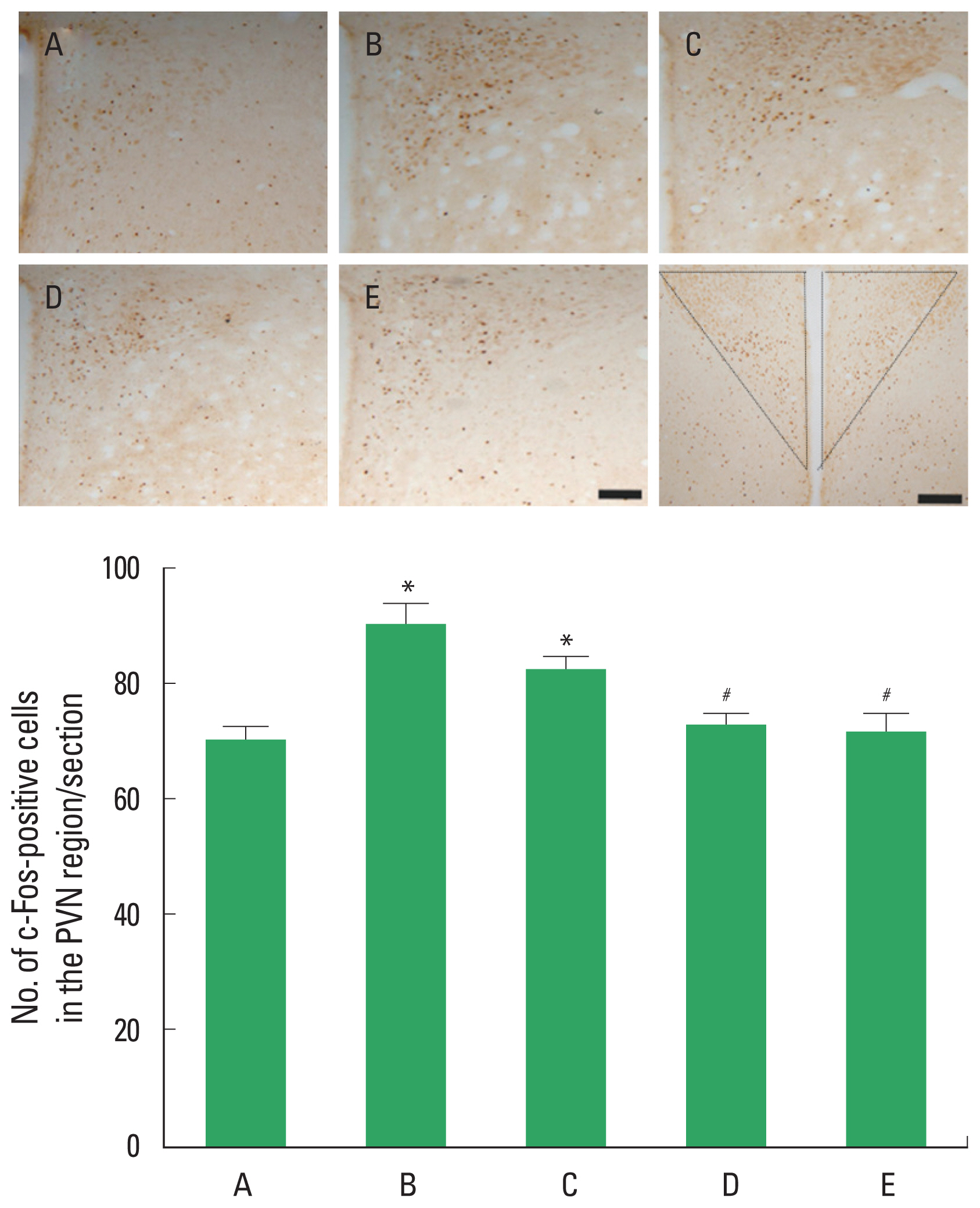
c-Fos expression in the PVN
c-Fos expressions in the PVN are presented in Fig. 2. The present results showed that the number of c-Fos-positive cell in the PVN was increased by SCI. Acupuncture at ST36-acupoint and EA treatment decreased the number of c-Fos-positive cells in the PVN of the SCI-induced rats.
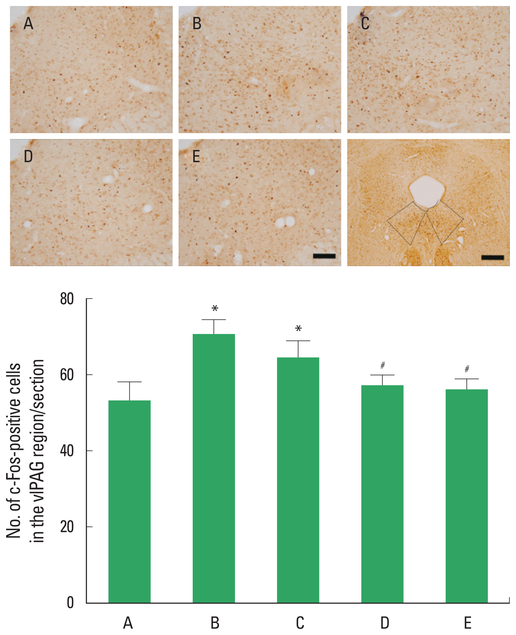
c-Fos expression in the vlPAG
c-Fos expressions in the vlPAG are presented in Fig. 3. The present results showed that the number of c-Fos-positive cell in the vlPAG was increased by SCI. Acupuncture at ST36-acupoint and EA treatment decreased the number of c-Fos-positive cells in the vlPAG of the SCI-induced rats.
NF-200 expression in the sciatic nerve
NF-200 expressions in the sciatic nerve analyzed by western blotting are presented in Fig. 4. The present results showed that NF-200 expression in the sciatic nerve was decreased by SCI. Acupuncture and EA treatment increased NF-200 expression in the sciatic nerve of the SCI-induced rats. EA showed most potent increasing effect on NF-200 expression.
BDNF expression in the sciatic nerve
BDNF expressions in the sciatic nerve analyzed by western blotting are presented in Fig. 5. The present results showed that BDNF expression in the sciatic nerve was increased by SCI. Acupuncture and EA treatment decreased BDNF expression in the sciatic nerve of the SCI-induced rats. EA showed most potent deceasing effect on NF-200 expression.
DISCUSSION
The SFI is widely conducted to evaluate functional walking performance after peripheral nerve lesion, since it integrates sensory and motor fiber on specialized pattern of movement (Dinh et al., 2009). EA relieves the behavioral signs of neuropathic pain (Gim et al., 2011).
In the present study, SCI showed characteristic gait change with decrease of the SFI value. The rats in the EA-treated group showed increased SFI value. These results indicate that EA treatment improved locomotor function following SCI.
Nociceptive information reaches to the brain from the peripheral injury through ascending neuronal pathways (Vermeulen et al., 2014). c-Fos is commonly used to indicate activation of neurons in the brain induced by external inputs. c-Fos is used as a sensitive marker of the neural activation following sciatic nerve ligation (Nishimori et al., 2002). de Medeiros et al. (2003) demonstrated the EA exerted analgesic effect and influenced c-Fos expression in the rats with peripheral neuropathic pain. Increased c-Fos expression represents peripheral nerve activation (Han et al., 2017; Ko et al., 2016).
In the present study, c-Fos expression in the vlPAG and PVN was increased following SCI, while EA treatment suppressed SCI-induced c-Fos expression in the vlPAG and PVN. The present results indicate that neurons in the vlPAG and PVN were activated by SCI. In contrast, EA suppressed noxious stimulation on sciatic nerve, resulting in decrease of neuronal activation in the vlPAG and PVN.
Another important characteristic of nerve damage is NF activation. NF is the intermediate filament of never cells and NF is abundant in axonal process. NF is involved in the axonal transport and maintenance of cell shape as cytoskeletal components (Park et al., 2013), thus, NF activation is suppressed in the nerve system by various damages. Decreased immunoreactivity of NF-200 was observed in the rats with sciatic nerve ligation, in contrast, decrease of hypersensitivity increased axonal NF-200 immunoreactivity (Tomassoni et al., 2018).
In the present study, NF-200 expression in the sciatic nerve was decreased by SCI, suggesting that NF-200 expression was disturbed by hypersensitivity caused by nerve injury. In contrast, EA treatment increased NF-200 expression. The present results indicate that EA alleviated SCI-induced pain sensitivity, and then resulted in enhancing of NF expression.
Peripheral inflammation increased BDNF synthesis in the dorsal root ganglion, which was mediated by nerve growth factor and an elevated anterograde transport of BDNF to axon terminals in the spinal dorsal horn (Cho et al., 1997). Following nerve injury, the expression of BDNF in the superficial dorsal horn was increased (Kim et al., 2001). BDNF may play a critical role in synaptic plasticity in nociceptive signaling and regeneration (Kim et al., 2017; Sparg et al., 2004). Also Huang et al. (2004) reported EA accelerates nerve regeneration and functional recovery in delayed peripheral nerve injury in rats. BDNF is closely implicated in new neuronal generation (Hwang et al., 2016). However, the protective effects of neurotrophic factors on nerves have often controversy (Zhao et al., 2015). Lee et al. (2018) demonstrated that BDNF expression in the sciatic nerve was increased by SCI, in contrast, treatment with diosgenin suppressed BDNF expression.
In the present study, BDNF expression in the sciatic nerve was increased by SCI, suggesting that injury to the sciatic nerve induced BDNF expression for its regeneration. In contrast, EA treatment suppressed SCI-induced BDNF expression, suggesting that EA facilitated regeneration of injury to the sciatic nerve, thus over-expression of BDNF was suppressed.
In the present results, acupuncture and EA showed effectiveness on functional recovery from SCI. Of these, EA showed more potent effectiveness on functional recovery from SCI.