INTRODUCTION
Gossypiboma literally means retained surgical sponge. Gossypiboma is derived from Latin word “gossypium,” which means “cotton” and “boma” in Kiswahili, which means “place of concealment”. Most often found in abdominal and pelvic cavities, retained gauze can cause a large variety of symptoms, including pain, palpable mass, fever, constipation, ileus, and fistulas (Wan et al., 2009). Two usual responses to retained surgical gauze are exudative reaction leading to abscess formation or fluid collection, and an aseptic fibrinous reaction resulting in adhesion, encapsulation, and eventual formation of granulomas of various sizes (Wan et al., 2004). The time of presentation may range from early post-operative period to several decades later. A correct diagnosis is difficult and can be made in only one-third of cases. Plain abdominal radiography, ultrasonography (USG), computed tomography (CT) scan and magnetic resonance imaging (MRI) are the diagnostic modalities available (Jason et al., 1979; Mochizuki et al., 1992; Moyle et al., 1996; Overton and Stone, 1968). A minimally invasive technique such as laparoscopy is a good diagnostic and therapeutic modality in cases without a definitive preoperative diagnosis. We report a case of retained surgical gauze diagnosed and removed by laparoscopic surgery, 24 yr after the Cesarean delivery.
MATERIALS AND METHODS
38-year-old athlete female patient admitted for a suspected pelvic mass, found on abdominal CT that was conducted during evaluation of lower quadrant (RLQ) pain. She felt intermittent pain in RLQ area for about one month. There was no history of abdominal pain before about 1 month ago. She had undergone cesarean delivery 24 yr earlier. She has no other history of operation. The patient has been doing well without complaints after cesarean delivery. She started to feel abdominal pain about 1 month ago.
On clinical examination, the patient was in good condition and her vital signs were normal. Her abdominal was not painful, and nearly fist-sized hard mass was palpitated on right side of uterus. All blood tests and urine analysis were within normal range. Ultrasonography revealed suspicious pelvic mass sized about 8 cm and it looked like subserosal myoma or right ovarian fibroma.
RESULTS
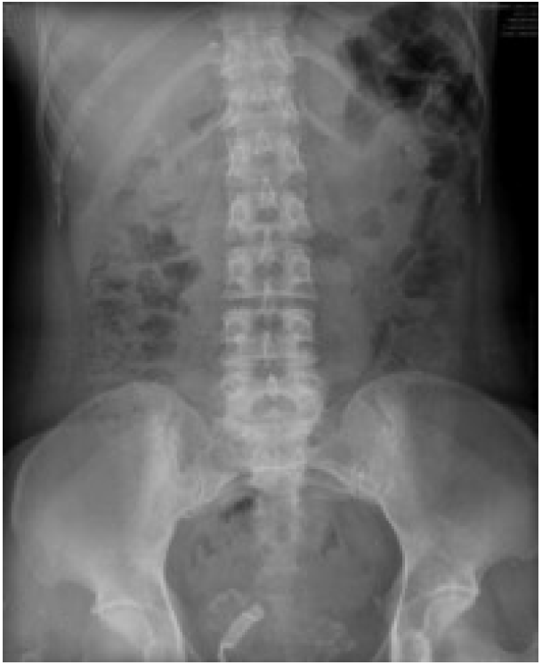
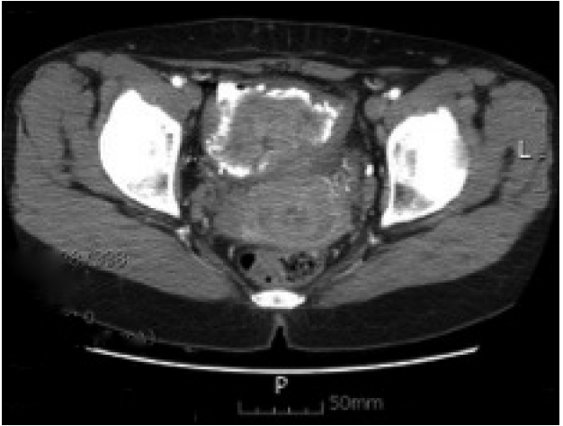
After admission plain abdominal radiography was performed, and irregular calcification suggesting tab-gauze in pelvis was found (Fig. 1). Contrast enhanced abdominal computerized tomography was also revealed that 9 cm sized mass lesion with irregular calcifications was seen at the anterior aspect to the uterus. It was not enhanced at all. It suggested gossypiboma probably tab-gauze (Fig. 2).
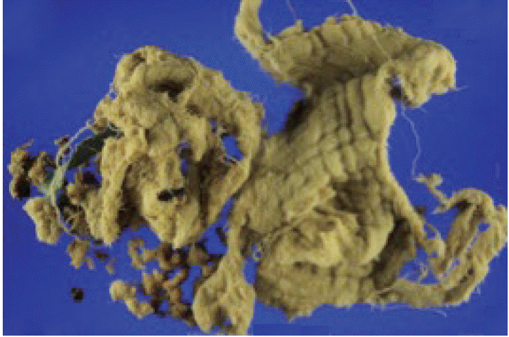
With the high index of suspicion, diagnostic laparoscopy was done. The anterior surface of uterus is conglomerated with pelvic mass. The pelvic mass was looked like tab-gauze and conglomerated with omentum, uterus and bladder (Fig. 3). Dissecting the mass was very careful, but because of severe adherence, bladder wall was lacerated about 1cm. The gauze was removed and bladder repair was done. A foley cathter was inserted during an admission period and the patient was discharged on 7th postoperative day keeping foley cathter. The foley catheter was removed on 15th postoperative day after cystography done.
DISCUSSION
Retained surgical sponge occurs at a frequency of one per 100–3,000 operations (Kaiser et al., 1996). Gossypiboma is a rare but preventable condition associated with many legal implications. The presentation may be acute or delayed just like this case. Radiologic investigations like USG, CT, and MRI can give a fair estimate of the diagnosis. In our case, we can have high index of suspicion by abdominal CT. But ambiguous clinical presentation in which a definite diagnosis is not available, laparoscopic intervention provides a diagnosis as well as simultaneous treatment. For this reason in these rare cases laparoscopy has an advantage for such rare presentations to achieve both diagnosis and treatment.
Gossypiboma always carries some medico-legal implications. The presence of a foreign body inside the patient is an avoidable problem and the patient may litigate the responsible surgeon. Moreover, gossypiboma may be misdiagnosed as a malignant tumor and lead to unnecessary invasive diagnostic procedures or extensive extirpative surgery which may result in further complications (Zbar et al., 1998).
Prevention is the best treatment. All surgical textile materials and instruments should be counted once at the start and twice at the conclusion of surgical procedures. However, counts are not always sufficient. It has been reported that most cases of gossypiboma occur in presence of normal pack counts (Rappaport and Haynes, 1990). Hurried counts that may occur in emergency operations or an unexpected change in procedure might also contribute to increase the incidence of gossypiboma.
When there are changes in theater personnel, the mistake is more likely. High body mass index of patients has been given as another risk factor (Gawande et al., 2003). On the other hand, when the accuracy of final count is in doubt, intraoperative radiologic screening may detect any retained surgical textile material impregnated with a radio-opaque marker. Moreover, Gawande et al. (2003) suggested the routine radiographic screening of high-risk patients before they leave the operating room even when counts are documented as correct. At the time of any operation, the operation field should be thoroughly examined for retained surgical textile materials and instruments, particularly where packs have been employed remotely from the site of main surgery.
Even though the surgeon carries the major responsibility, this problem can only be overcome if all the members in an operation team work together meticulously as concluded by Schelhaas and Mastboom (2002).