INTRODUCTION
Patellofemoral pain is characterised by anterior or retropatellar pain associated with activities that load the patellofemoral joint, such as ascending or descending stairs, squatting, running and kneeling. Patellofemoral pain syndrome (PFPS) is the most common complaint affecting the knee (Wilson, 2007). 18.5–31% in young adults complained of knee pain and 50% of the non-specific knee pain accounted PFPS (Rathleff et al., 2012). The factors causing PFPS include weakness and imbalance of Quadriceps muscle, malalignment of lower limb, stiffness of soft tissue, increase in Q-angle of quadriceps muscle, and overuse and abnormal movement of hip joint (Bolgla and Boling, 2011). Particularly if there is an imbalance of quadriceps muscle of thigh and a shortening of the lateral support attached to the lateral side of knee joint, patellar is an occurrence of tilting to the lateral side in the knee joint, which is a common symptom of PFPS (Lun et al., 2005). PFPS has a risk of developing into chronic diseases such as chondromalacia or arthritis and so proper care and treatment is needed (Jeong and Kim, 2010).
Vastus medialis and vastus lateralis of quadriceps muscles are countervailing to adjust the location of knee joint and finally enhance the dynamic efficiency in knee function (Tang et al., 2001). Especially, vastus mediali has been suggested to act as a dynamic medial stabilizer (Gilleard et al., 1998; McConell, 1986), PFPS appear to decrease vastus medialis, that it may increase the lateral pull of the patella and reduce function at the knee joint (Baker et al, 2002; Chrisou, 2004). The patella malalignment changes the normal mechanism of knee extending, which brings about pathological changes of patellofemoral joint (Smith et al., 2009). To recover the mechanism of patellofemoral joint extending, locating an exact location of knee joint is important to increase the activity of vastus medialis and recover the start time, and more important to treat the patient with PFPS (Cowan et al., 2002). The conservative treatments of PFPS include taping to adjust the misalignment of lower limbs, stretching of soft tissue, strength of lower limbs, neuromuscular control training of quadriceps muscle of thigh, bio-feedback exercise (Aminaka and Gribble, 2008).
Corrective taping of the patella has since become a commonly used treatment for PFPS (Herrington, 2000). McConnell taping has devised a classification to describe abnormal patellar alignment. The four main malalignments include excessive lateral glide; excessive lateral tilt; excessive posterior tilt of the inferior pole and excessive rotation (McConnell, 1986, 1996). McConnell taping is designed to correct these malalignments and has four basic components, medial glide, medial tilt, anterior tilt and rotation. It is suggested that taping affects the tracking of the patella, facilitating its centralization within the trochlear groove (McConnell, 2002). If we look at the application of patellar taping, Christou (2004) applied patellar taping to normal subject and patient with PFPS and used isokinetic measuring instrument to see how active the muscle activity of vastus medialis and that of vastus lateralis is in terms of knee angle. As a result, the activity of vastus medialis increased, but that of vastus lateralis decreased in patients with PFPS. On the other hand, the activity of vastus medialis decreased, but that of vastus lateralis increased in normal person. Gilleard et al. (1998) investigated the effect of patellar taping on the timing of vastus medialis and vastus lateralis muscle activity in 14 female subjects with PFPS during stair ambulation with and without patellar taping. As a result, the onset of vastus medialis EMG activity compared with vastus lateralis occurred earlier in the knee movement with patellar taping than without taping during a step-up and step-down test. The authors discussed the possibility that patellar taping may enhance the onset of vastus medialis activity, which may result in improved patellar tracking (Gilleard et al., 1998). Many several studies showed that patellar taping is effective to reduce pain, increase the muscular strength of quadriceps muscle of thigh (Crossley et al., 2001), improve the neuromuscular mobilization, and correct the muscular contracture start time of vastus medialis against vastus lateralis (Cowan et al., 2002). However, most studies were restricted to examining pain or muscle activity of the quadriceps femoris muscle during simple actions and those concerning taping effects of the patella during functional motions including knee bending have been insufficient.
This study was to investigate the effects of muscle activity of quadriceps in the functional activity of squatting with and without McConnell taping.
MATERIALS AND METHODS
Subjects
The number of participants in this study is 16 patients (8 Female, 8 Male) with knee anterior pain. The subjects of this study were patients with knee joint pain who visited P hospital located in Daegu Metropolitan City during October 2012. The subjects were limited to a person less than 80 points in funtional question-naries (Kujala Patellofemoral Score) (Barby and Kevin, 2009).
The average age of subjects was 31.69±4.17 yr, average height was 167.51±8.24 cm, and average weight was 61.82±10.90 kg. After explaining about the purpose and content of this study prior to experiment to each and every participant, they are given a consent form. The requirements for experimental subjects are as follows: First, they should be less than 80 points in Kujala Patellofemoral Score. Second, they should be no history of hip joint, ankle, and foot joint dysfunction. Third, they should have no difficulty in squatting down. Fourth, they should have no orthopedic surgery history in lower extremity.
Experimental procedure
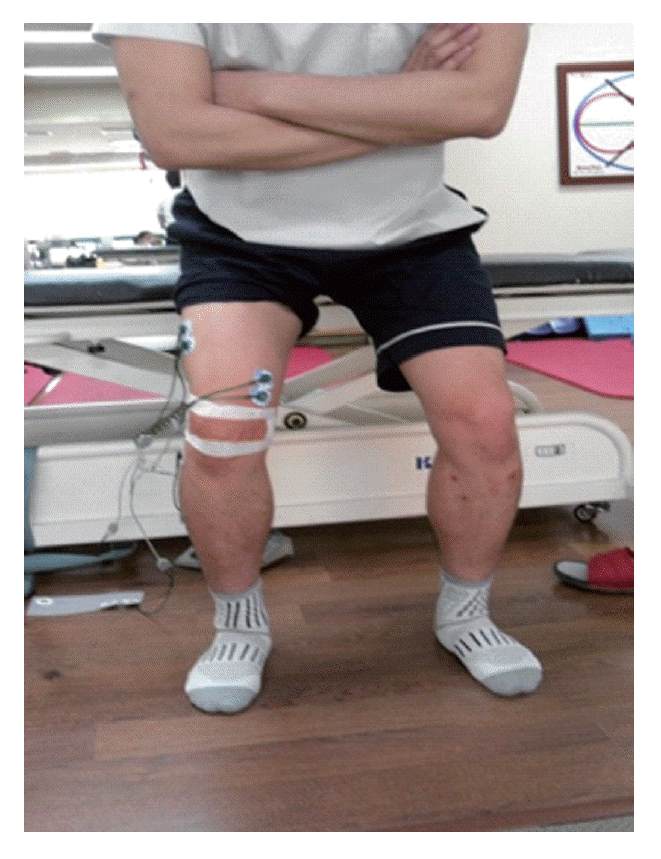
Sixteen participants squat in the following three conditions: no-taping, placebo taping with no tilting of knee joint, and patellar taping with medial tilting of knee joint. In no-taping, they squat with no taping attached. In placebo taping, they are taped at the same place as patellar taping without inducing the medial attraction of knee joint. In patellar taping, McConnell taping (McConnell, 1986) is applied, the knee joint is pushed toward the inside to induce medial attraction, and then they are taped before they squat. The advenced fixing tape (Endura-FIX, China) is attached to prevent the skin slipping and then nonelastic tape (Endura-FIX, China) is attached on it. A skilled physical therapist with more than 10 years of experiences helped to tape it.
When squatting down, their two foot angle is 120% on the basis of their shoulder (Earl et al., 2001), their two arms folded in the front, and trunk stood up to avoid lumbar flexion. For the knee angle, a semi-squatting posture is adopted as it is widely common to patients with PFPS: 50° angle (Tang et al., 2001). Before exercise, 50° is measured and then a bar is installed so that each participant could participate in the experiment at the same angle. For the order of exercise, the order is distributed randomly by drawing cuts. When doing each movement, they should keep for 5 sec and rest for 30 sec, which is repeated three times. After exercise, tape is replaced by another one. And then they squat in another condition (Fig. 1).
EMG and data processing
For data collection, EMG Myosystem 1200 (Noraxon Inc., USA) System is used to measure the muscle activity of vastus medialis and vastus lateralis according to the taping. The attachment location of vastus medialis is 55° in a vertical line from the upper and inner point of the knee joint: 4 cm upper and 3 cm inner from the knee joint. That of vastus lateralis is 15° from the upper line of knee: 10 cm upper and 6–8 cm outer. The distance between electrodes is constant at 2 cm intervals. The single reference electrode is attached to knee joint. Before electrode attachment, skin is cleaned with an alcohol swab and shaved to reduce the skin resistance to less than 5 kΩ (Cowan et al., 2002). To obtain the maximum torque of each muscle, maximal voluntary isometric contraction (MVIC) of vastus medialis and vastus lateralis is obtained at 60° knee joint in the squatting posture (Dixon and Howe, 2007). In the analysis of muscle activity, MVIC is measured with the root mean square (RMS) measured when contracting for 5 sec from the knee muscle strength test (Kendall et al., 2005). After measuring each muscle activity in three different taping conditions, it is regarded as RMS and then each muscle activity for 3 sec in the middle is converted into a percentage (% MVICRMS) as in the below. And the ratio between vastus medialis and vastus lateralis is calculated with normalized muscle activity value in each and different taping condition.
Data analysis
All measures from this experiment are statistically treated by SPSS ver 12.0 (ICC, Chicago, USA). One-way repeated measured ANOVA is used to see if there is a difference between the measured muscle activity of vastus medialis and vastus lateralis and the ratio between the measured vastus medialis and vastus lateralis in three different conditions: no taping, patellar taping, and placebo taping. In post-test for the significant difference, pair-comparison is conducted with Bonferroni’s correction. The significant level for statistical significance testing is at α=0.05.
RESULTS
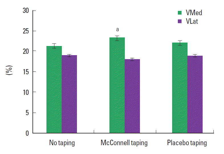
The average muscle activity of vastus medialis is 21.36±11.70% in no taping, 23.33±12.08%, in McConnell taping, and 22.10 ±10.64% in placebo taping. The average muscle activity of vastus lateralis is 19.02±9.08% in no taping, 18.08±7.34% in McConnell taping, and 18.92±8.96% in placebo taping. The activity ratio between vastus medialis and vastus lateralis is 1.14±0.40% in no taping, 1.29±0.42% in McConnell taping, and 1.20±0.36% in placebo taping.
In the comparison of the muscle activity between vastus medialis and vastus lateralis, the activity is significantly reduced in McConnell taping (P<0.05). Also, in the comparison of the activity ratio between vastus medialis and vastus lateralis, the activity is significantly reduced in McConnell taping (P<0.05). On the other hand, placebo taping is not statistically significant in the muscle activity between vastus medialis and vastus lateralis and the activity ratio between vastus medialis and vastus lateralis (P>0.05) (Table 1) (Fig. 2).
DISCUSSION
The lateral tilting of patella is caused by the imbalance of vastus medialis and vastus lateralis. The shortening of the lateral support causes the relatively at weakness vastus medialis and so does not contribute to the dynamic medial stabilization (a major action of vastus medialis), thus resulting in abnormal movement of knee joint (McConnell, 2007). In McConnell taping, the use of inelastic tape is known to be effective to induce the medial attraction of lateral tilting knee joint, reduce pain with fixation, affect the functional activity, and change the start time of vastus medialis associated with vastus lateralis and reeducate the nerve root (Keet et al., 2007).
In this study, after McConnell taping to patellofemoral pain syndrom patients, they squat. The findings are that vastus medialis has the highest activity in placebo taping, followed by no-taping, McConnell taping and that the higher activity between vastus medialis and vastus lateralis indicates that McConnell taping may reduce the muscle activity; the values are significant. To see the similar previous studies to this founding, Evangelos and Christou (2004) applied McConnell taping to normal subject and patient with PFPS and used 30°/s isokinetic leg presses measuring instrument to see how active the muscle activity of vastus medialis and vastus lateralis, pain. The medial patella taping procedure reduced preceived pain (70%) and increased the activity of the vastus medialis and decreased the activity of vastus lateralis. In a study that examined maximal isometric muscle strength by applying patella taping, both concentric contraction and eccentric contraction increased (Herrington, 2001). According to the above studies, patella taping repositioned patellas in the trochlear groove, decreasing pressure in the patellofemoral joints, thereby effectively improving muscle strength of the quadriceps femoris muscle. And, McConnell was suggested, by using tape to correct patellar position, the vastus medialis is assisted in its efforts to resist the pull of the vastus lateralis and stabilize the patella (McConnell, 1996).
In this study, it is not statistically significant, but the muscle activity of vastus medialis and the activitiy ratio between vastus medialis and vastus lateralis increases numerically in placebo taping group compared to no-taping group. In a prior study, Christou (2004) noted that patella taping that was applied to patients with patellofemoral pain syndrome increased muscle activity of the vastus medialis and explained such change through decrease in pain of patellofemoral ligament and skin afferent stimulation. The result seems to be that taping promotes the tendon origin of vastus medialis and then afferent input is stimulated, and thus the increase of stimulation of α-motor neuron source increases the muscle activity.
In the present study, a group to which McConnell taping was applied for medial drawing saw muscle activity of the vastus medialis and vastus medialis (VMO): vastus lateralis (VL) activation ratio increase and the reason may be explained as follows. Cross-bridge formation by correcting the location of the patella and facilitation of muscle activity according to increase in skin afferent input is considered to have affected VMO activity.
This study is conducted in 16 adults in the late 20s and early 30s, has problems of not considering age variable and limitations in generalizing the finding due to the limited number of subjects. And a study on correlation between changes in pain and changes in muscle activity is considered to be necessary. Also, participants couldn’t take the same posture due to physical differences between individuals. More sophisticated equipment and optimal laboratory and apparatus are required and above problems should be supplemented.