INTRODUCTION
Although cancer survivorship is clearly increasing, there appears to be no improvement in the rate among older women over 70 years of age (Lundberg et al., 2022). The incidence of gynecological cancers has gradually increased (Benedet, 2000; Zhang et al., 2021). Cancer survivors experience considerable stress due to health concerns such as cancer cell metastasis and the sense of isolation they have experienced in the past (Tang et al., 2016). Long-term stress may lead to the rapid growth of cancer cells or metastasis of new cancer cells in patients who underwent treatment previously (Neeman and Ben-Eliyahu, 2013). Therefore, it is necessary to develop an intervention program that can help reduce the stress of survivors and an immune-enhancing therapy program capable of blocking the metastasis of new cancer cells.
Several studies have shown that the fragrance of phytoncides reduces stress and promotes immunocyte growth, including natural killer (NK) cells (Akram et al., 2020; Li et al., 2007; Vivier et al., 2008). Phytoncides are volatile substances that relieve stress and exert antibacterial effects when inhaled from the forests (Jo et al., 2021; Li et al., 2006; Li et al., 2007; Li et al., 2008), and they are most released from conifers, particularly pines, nut pins, and cypress trees. Essential oils from the Hinoki cypress and pine trees are primarily used in commercial products containing various terpenes (Jo et al., 2021; Lee et al., 2015), of which monoterpenes have adrenal cortex stimulation, antisepsis, sterilization, and antiviral effects. Tannin, the second most abundant component of phytoncides, has detoxification, hemostatic, and anti-inflammatory effects (Akram et al., 2020).
People have long believed that living in the woods improves the health of patients with cancer in Korea; however, the reason for this remains unclear. Although some researchers have argued that phytoncides activate the NK cells of patients with cancer to enhance immunocytes, others have suggested that phytoncides through the forests help reduce stress and the negative effects of chemotherapy (Tønnesen et al., 1987). Bodner et al. (1998) reported that when the level of cortisol increases, that of NK cell decreases, whereas when the level of cortisol decreases, that of NK cell increases. Tønnesen et al. (1987) reported that adrenaline produced an immediate increase in NK cell activity and a selective increase in circulating NK cells. Salleh (2008) also reported that NK cells respond sensitively to changes in cortisol or epinephrine in the blood.
The diverse experimental settings make it challenging to draw definitive conclusions from the findings of multiple previous studies regarding the effects of phytoncide fragrance on stress hormone responses and adaptations. That is, as reported in previous studies, stress hormones affect NK cells, and it can be said that phytoncide fragrance affects NK cells because it affects stress hormones. However, research conducted using phytoncide fragrance made from essential oils in urban areas is extremely rare, as research has only been conducted in external forest environments. Specifically, the mechanism by which the inhalation of the scent of phytoncides oil affects the endocrine system, the nervous system, and NK cells and subsets remains unclear. Thus, this study investigated the effects of phytoncide oil fragrance on the stress index, neuroendocrine concentration, and NK cell family in gynecological cancer survivors.
MATERIALS AND METHODS
Study design and participants
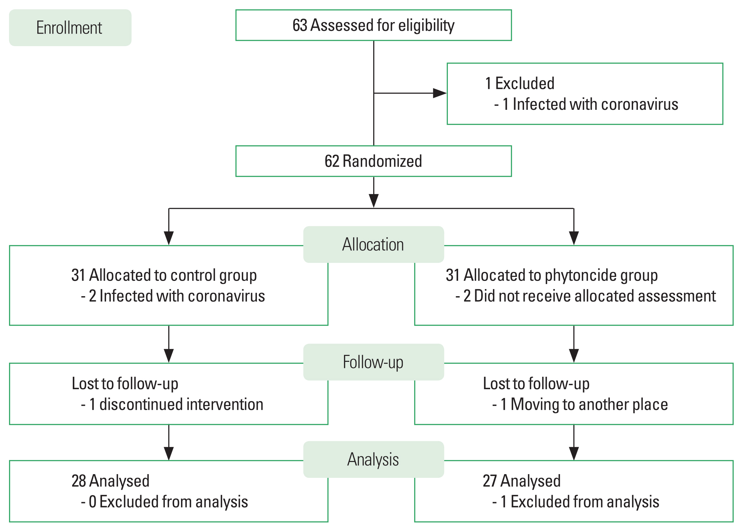
This prospective single-blinded randomized controlled trial was conducted in accordance with the Declaration of Helsinki and approved by the ethics committee (HS22-11-01). The study was registered with the Korean Clinical Research Information Service (KCT0008283). All the participants listened to the experimental procedures and provided informed consent before the study, and all data were acquired from November 2, 2022, to January 3, 2023. The participants were between 61 and 79 years old and gynecological cancer (cervical, ovarian, or breast cancers) survivors who were diagnosed between January 2016 and December 2020 and underwent surgery, radiation therapy, or chemotherapy, with a survival rate of less than 5 years. They were subjected to continuous management at Seoul Songdo Hospital, Korea. Patients with serious inflammation, osteoporosis, or psychological disorders were excluded. Although this study aimed to recruit 60 participants considering the dropout rate, 63 participants initially participated. Only 62 participants were included in the random classification because one person encountered the coronavirus disease. Participants were randomized into groups using the Research Randomizer program (Urbaniak and Plous, 2023); each participant was asked to draw a numbered ticket from a jar, where the number ranged from 1 to 62. Thirty-one participants each were assigned to the control group (CG) or phytoncide group (PTG). Some participants in both groups dropped out during the experiment (Fig. 1).
Intervention
Phytoncides oil (Chamaecyparis obtusa) was selected for olfactory stimulation (Raha et al., 2019), and an oil burner (Good Earth Lab, Seoul, Korea) made of cypress wood was used. The main oil components were terpenes, primarily monoterpenes and sesquiterpene (Lee et al., 2010). Three to five drops of phytoncides oil were dropped onto the burner to allow the wood to absorb and emit a subtle scent. The temperature and humidity of the closed laboratory space (approximately 66 m2) were maintained at 25°C–26°C and 50%–60%, respectively, to maintain the scent diffusion. The PTG meditated by lying down in a space scented with phytoncide for 1 hr a day, 5 days a week, for 8 weeks. The CG was allowed to meditate while lying down for the same time and period in a different place without phytoncide scent. The participants were classified according to their community areas to prevent communication between the groups.
Stress scale
This study used a 10-item version of the perceived stress scale (PSS) to identify stress levels (Cohen et al., 1983). PSS consisted of six positive items and four negative items. Each item was rated based on the past month on a Likert scale, which composed ‘never’ (0) to ‘very often’ (4) (Table 1). In scoring the measures, the positive items were reverse-scored and then aggregated (Golden-Kreutz, 2004). The score ranges 0–13, 14–26, and 27–40 were considered low, moderate, and high perceived stress, respectively. The reliability coefficient, assessed using Cronbach alpha, was 0.913. We conducted a structural model analysis to confirm if the potential variables on the stress scale explain the observed variables.
Body composition and controlled variables measures
The participants were asked to limit their food and caffeine intake before the test, urinate and not exercise the day before the test. All participants removed the metals attached to their bodies, and the body composition of each participant was measured using an InBody 770 (Biospace Co. Ltd., Seoul, Korea) after their height was measured. Additionally, daily diet by CAN-Pro 5.0 (The Korean Nutrition Society, 2023) and physical activity level by the International Physical Activity Questionnaire (Booth, 2000) were recorded. Calorie intake and expenditure through physical activity were checked daily, recorded on a computer log, and averaged at the end of the month.
Autonomic nervous system measures
All participants were asked to abstain from working out, ingest food 3 hr, and take a break 30 min before the test to obtain a measurement of heart rate variability (HRV). The uBioMacpa (Biosensecreative Co. Ltd., Seoul, Korea) was used for HRV test. Frequency analysis of HRV provided data on the following HRV components: high frequency and low frequency (Lane et al., 2009). Since it was clear that high frequency is an activity indicator of the parasympathetic nervous system (PNS), the prevailing view was that low frequency mainly reflects sympathetic nervous system (SNS) activity (White and Raven, 2014).
Stress hormones and immunocytes measures
The participants had fasted for at least 10 hr and 20 mL of blood was taken from the antecubital vein. It was centrifuged at 3,000 rpm for 15 min and stored in a −80°C freezer until analysis. Whole blood samples were used for automated differential blood cell count and fluorescence-activated cell sorting (FACS) analyses. Peripheral blood samples were collected from heparinized collection tubes and ethylenediaminetetraacetic acid (EDTA) tubes. Cortisol levels were measured using an enzyme-linked immunospecific assay (ELISA) kit (DRG Instruments GmbH, Hamburg, Germany). Epinephrine levels were analyzed using a high-performance liquid chromatography with a plasma catecholamine ELISA kit (Labor Diagnostika Nord, Nordhorn, Germany). EDTA-anticoagulant blood samples were tested using Sysmex XN-550 (Sysmex Corp., Kobe, Japan). Particularly, the samples were stored at room temperature for 4 hr before the test. A melted whole-blood technique with a maximum of 8-color staining was used for flow cytometry and antibody staining. Notably, 50 μL of blood was with anti-human antibodies against anti-CD56, anti-CD3, anti-CD314 (NKG2D), anti-CD158b (KIR2DL3) (BioLegend, San Diego, CA, USA), and isotype controls (Martinez-Borra and Khakoo, 2008). Moreover, T-cell immunoreceptors with immunoglobulin (Ig) and immunoreceptor tyrosine-based inhibitory motif (TIGIT) and lymphocyte function-associated antigen-1 (LFA-1) were analyzed. After incubation for 30 min at room temperature in the dark, the erythrocyte was lysed by adding 500 μL of FACS lysing solution to each test tube for 15 min. Upon completion of staining, the cells were analyzed using FACS Canto II (BD Biosciences, San Jose, CA, USA) and the FlowJo program (Treestar, Ashland, OR, USA) and the results are presented as percentages.
Statistical analyses
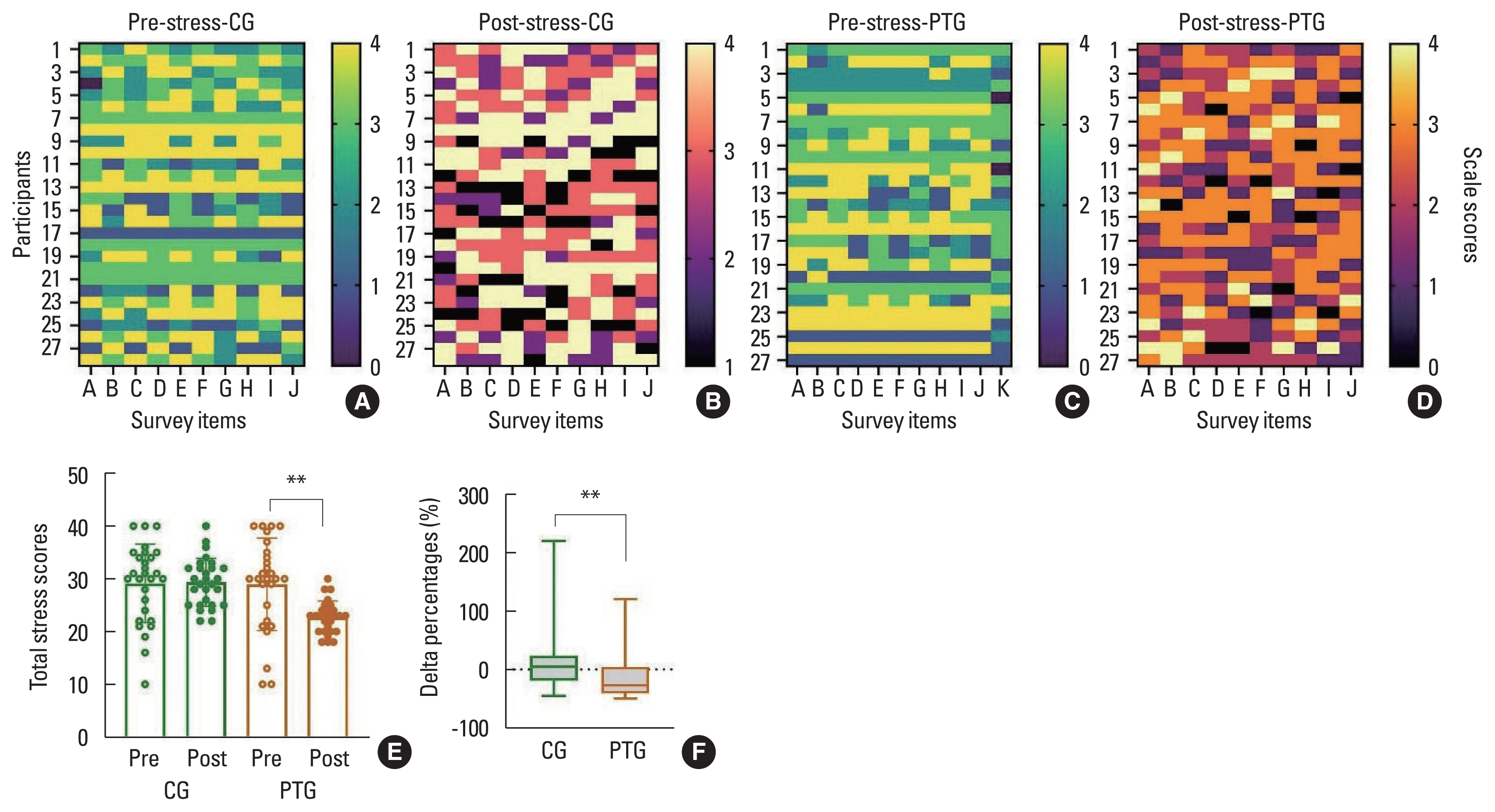
Utilizing G·Power program, the sample size was obtained based on an a priori effect size (0.25) of f 2 (V), α error probability (0.05), power (0.95), two groups, and two measurements (Faul et al., 2007), and the total sample size was 54. The software used in this study for analyses were SPSS and AMOS (ver. 25; IBM Corp., Armonk, NY, USA) and GraphPad Prism 9.5.1 (Graph Pad Software, San Diego, CA, USA). Normal data distribution was estimated using D’Agostino and Pearson omnibus tests. The stress scale showed scores for 10 questions (A to J) for each participant in each group using a heatmap. Confirmatory factor analysis was conducted to verify the validity of the observed variables constituting the latent variables of the stress questionnaire. This study analyzed the prescores of the variables among the groups using the Mann–Whitney U-test. Two-way analysis of variance and Wilcoxon signed-rank tests were used to compare the results between groups and times. A delta percentage (%) analysis was also conducted, and effect size (η2) was computed according to Cohen d (Cohen, 1992). All data are presented as mean±standard deviation and analyzed based on P≤0.05.
RESULTS
Demographic and clinical characteristics
As shown in Table 2, there were no significant differences in age, body composition components, demographic properties, surgical stages, and therapeutic methods between the groups before the experiment.
Specifically, the calorie intake of CG at week 4 was 1,608.36± 176.31 kcal/day, whereas the calorie intake of PTG was 1,588.85± 167.51 kcal/day, showing no statistically significant difference (Z= −0.338; P=0.736, η2=0.003). Even at week 8, the calorie intake of CG was 1,752.29±249.99 kcal/day, whereas the calorie intake of PTG was 1,725.41±141.00 kcal/day, showing no significant difference (Z=−1.207; P=0.228, η2=0.004) between the groups. The calorie consumption of CG was 534.21±62.79 kcal/day, whereas that of PTG was 575.37±10.04 kcal/day at week 4, showing no statistically significant difference (Z=−1.672; P=0.095, η2= 0.103). At week 8, the average calorie consumption of CG was 583.82±33.43 kcal/day, whereas that of PTG was 583.30±32.42 kcal/day, showing no significant difference (Z=−0.110; P=0.913, η2=0.007) between the groups.
Stress scale and implication of phytoncide fragrance on stress level
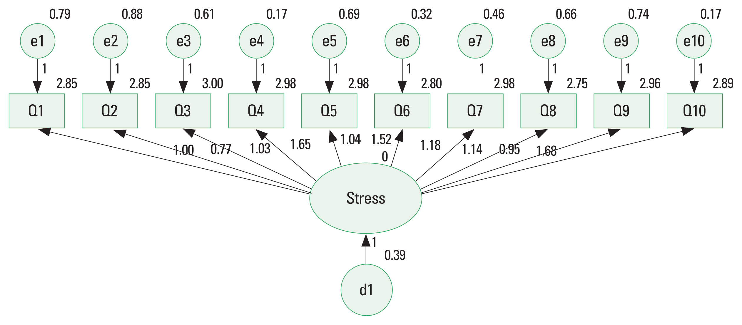
In the fit of the assessing model, the χ2 (df=35) value was 170.58 (P<0.001). Since the χ2 value was greatly affected by the sample size and complexity of the model, other goodness-of-fit indices were identified. The results showed that the comparative fit index=0.905, Tucker–Lewis index=0.914, and root mean square error of approximation=0.036 were good, and the test model was found to be suitable (Fig. 2). All factor loadings were found to have a significant positive effect in the confirmatory factor analysis except for Q3 and Q5 (Table 3).
Fig. 3 shows the stress levels of participants before and after the experiment based on the stress scale. Before the experiment, the total stress levels in CG and PTG were 29.14±7.46 and 28.96± 8.77, respectively; however, that of CG increased to 29.36±4.53 (Z=−0.048, P=0.962), while that of PTG decreased to 22.70± 3.10 (Z=−2.921, P=0.003) after 8 weeks (Fig. 3E). The Δ% total stress levels of CG was 10.89±49.59%, while that of PTG was −9.31%±45.98%, indicating a significant difference between the groups (Z=−2.568; P=0.010, η2=0.210) (Fig. 3F).
Implication of phytoncide fragrance on autonomic nervous activity
As shown in Fig. 4A, the SNS of CG was 7.16±0.85 before the experiment and increased to 7.38±1.00 (Z=−1.166, P=0.243) after 8 weeks. The SNS of PTG was 7.34±0.62 before intervention and decreased to 6.07±0.82 (Z=−3.965, P=0.001) after 8 weeks. The Δ% of CG was 3.82%±14.7%, while that of PTG was −16.75%±13.25% (P=0.001, η2=0.359). These results showed significant differences and changes in the group (P=0.002, η2= 0.166), time (P=0.001, η2=0.205), and interaction (P=0.001, η2=0.339).
The PNS of CG was 3.72±0.49 and decreased to 3.39±0.37 (Z=−3.737, P=0.001) after the experiment, as shown in Fig. 4B. Meanwhile, the PNS of PTG increased from 3.56±0.45 to 4.10± 0.47 (Z=−3.245, P=0.001) after 8 weeks. The Δ% of CG was −8.31%±10.05%, while that of PTG was 16.93%±20.42% (P= 0.001, η2=0.392). These results showed significant differences and changes in the group (P=0.004, η2=0.143) and interaction (P=0.001, η2=0.379).
The implication of phytoncide fragrance on stress hormones
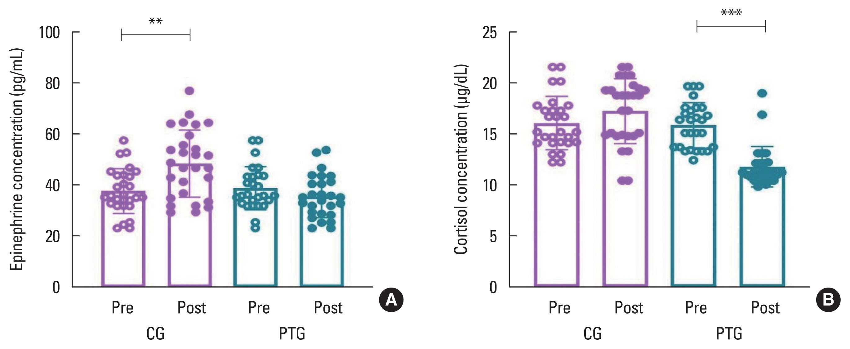
As shown in Fig. 5A, the epinephrine of CG was 37.90±8.84 pg/mL before the experiment and increased to 48.68±13.25 pg/mL (Z=−3.131, P=0.002) after it. Conversely, the epinephrine of PTG was 39.03±8.53 pg/mL before intervention and decreased to 35.74±8.35 pg/mL (Z=−1.412, P=0.158) after 8 weeks. The Δ% of CG was 34.31%±46.46%, while that of PTG was −5.29%± 25.5% (P=0.001, η2=0.223). These results showed significant differences and changes in the group (P=0.005, η2=0.139), time (P=0.040, η2=0.077), and interaction (P=0.001, η2=0.228).
The cortisol of CG was 16.18±2.63 μg/dL before the experiment and increased to 17.36±3.19 μg/dL (Z=−1.139, P=0.255) after 8 weeks, as shown in Fig. 5B. In contrast, the cortisol of PTG was 15.96±2.22 μg/dL before intervention and decreased to 11.87± 2.01 μg/dL (Z=−4.474, P=0.001) after the experiment. The Δ% of CG was 9.0%±23.62%, while that of PTG was −24.94%± 11.62% (P=0.001, η2=0.460). These results showed significant differences and changes in the group (P=0.001, η2=0.330), time (P=0.001, η2=0.193), and interaction (P=0.001, η2=0.441).
The implication of phytoncide fragrance on immunocytes
Table 4 presents NK cells and their subsets showed significant positive changes except for CD56bright NK cell. Among them, KIR2DL3 showed an increased tendency in CG and a decreased change in PTG. The remaining NK cells, subsets, and receptors showed decreased changes in the CG and increased changes in the PTG. Regarding the rate of change, the total NK cell of CG was 0.61%±37.1%, while that of PTG was 23.32%±32.94% (P= 0.019, η2=0.098). Moreover, the CD56dim NK Δ% in CG was −2.43%±3.94%, while that of PTG was 1.45%±4.02% (P=0.001, η2=0.198). The NKG2D Δ% in CG was −1.97%±4.22%, while that of PTG was 3.02%±3.24% (P=0.001, η2=0.311). The KIR2DL3 Δ% in CG and PTG were 8.36%±21.82% and −7.56%± 19.87%, respectively, indicating a significant difference between groups (P=0.006, η2=0.131). The TIGIT Δ% in CG and PTG were −4.33%±23.6% and 23.26%±25.69%, respectively (P= 0.001, η2=0.245). The perforin Δ% in CG and PTG were −6.17%± 7.30% and 3.21%±4.84%, demonstrating a significant difference between groups (P=0.001, η2=0.372). Finally, the LFA-1+NK Δ% in CG and PTG were −3.56%±5.68% and 6.08%±10.02%, representing a significant difference between groups (P=0.001, η2=0.269).
DISCUSSION
This study confirmed that gynecological cancer survivors had moderate-to-high stress levels that decreased after phytoncide intervention. In addition, the study discovered that cancer survivors who were exposed to phytoncide fragrance experienced a beneficial impact on their stress hormones, as it suppressed the SNS and activated the PNS. Furthermore, a decrease in stress hormones was found to enhance NK cells and their subsets after being treated with phytoncide.
A fragrance is a volatile chemical compound that is perceived through olfaction (Ko et al., 2021; Woo and Lee, 2020; Zarzo, 2007). Olfactory receptors detect molecules and produce emotional responses that carry signals to the brain (Royet and Plailly, 2004). The main components of phytoncides are terpenes, which have rejuvenating effects in humans (Houdkova et al., 2020). Ikei et al. (2015) reported that essential oils extracted from phytoncides could be used to treat stress. Similarly, this study found that the stress level of the PTG decreased by approximately 9% after phytoncide intervention; this stress reduction was due to changes in the levels of neuroendocrine substances. Among the results of this study, the SNS activity was significantly decreased in the PTG (~−17%), while the PNS activity was significantly increased in the PTG (~17%). It is estimated that the effect of the phytoncide scent on stress is positive when it is absorbed into the body (Li et al., 2008). Phytoncide reduces stress by stimulating the olfactory bulb, which activates the autonomic nerve system (Angelucci et al., 2014). However, few studies have examined which hormones are affected by the autonomic nerve system and immunocytes (Segerstrom and Miller, 2004). Therefore, this study aimed to fill this gap, and it was found that phytoncide scent suppressed the SNS while activating the PNS. According to the results of this study, epinephrine and cortisol levels tended to decrease after applying phytoncide fragrance, though cortisol levels reduced more than epinephrine levels. Notably, the epinephrine concentration Δ% of PTG decreased by ~5%, while that of CG increased by ~34% after 8 weeks. The cortisol concentration Δ% of the PTG decreased by ~25%, while that of CG increased by ~9% after 8 weeks. Based on the effects, the phytoncide scent caused a positive change in the cortisol hormone in gynecological cancer survivors.
NK cells and their subsets were altered by stress hormones, as verified in this study, suggesting that the reduction of epinephrine and cortisol activates NK cells (Bodner et al., 1998; Tønnesen et al., 1987). Li et al. (2006) stated that the target cells are killed when phytoncides are used to treat an NK cell line. They also suggested that phytoncides increase NK cell activity by inhibiting cortisol. Moreover, NK cells activated by increased phytoncide levels are related to increased perforin, granzyme B, and granulin (Li et al., 2006). Thus, NK cells were influenced by their subsets. Specifically, the expression intensity of CD56 further divided NK cells into two subsets (CD56dim and CD56bright NK cells) (Phan et al., 2017). While CD56dim exhibited cytotoxicity, CD56bright showed less cytotoxicity but regulated immunocytes by secreting various cytokines (Raulet, 2003). Among these two subsets, this study found that CD56dim NK, rather than CD56bright NK, had a greater effect on the phytoncide intervention.
Typically, NKG2D+NK receptor detects UL16-binding proteins and major histocompatibility complex (MHC) class-I-chain-related proteins, which are intracellular molecules whose expression is increased during cancer (Martinez-Borra and Khakoo, 2008). In contrast, the inhibitory receptors of NK cells measure the presence or absence of intracellular molecules that are constitutively expressed on the surfaces of target cells (Long, 2008). Specifically, human KIR is an MHC class I-specific inhibitory receptor expressed in NK cells (Di Santo, 2008). This study showed that NKG2D+NK increased, while KIR2DL3+NK of the PTG decreased. These results suggest that phytoncides affect immunocyte receptors, even if provided as an aroma compound, as the blood flow of the chemical function in the tissues is improved. In addition, TIGIT, perforin, and LFA-1+NK cell levels decreased in the CG, while those increased in the PTG. Specifically, genetic ablation of TIGIT has been reported to enhance the NK cell-killing function (Urlaub et al., 2017). One of the most important weapons of killer T cells is perforin, a protein that pierces the outer membrane of target cells and causes cell necrosis (Trapani and Smyth, 2002). Moreover, LFA-1 was critical for the efficient activation and cytotoxicity of NK cells (Urlaub et al., 2017). As known, the subsets of NK cells are more mature and have higher cytotoxic potential to activate LFA-1 more.
This study observed that NK cells, including their subsets and receptors, were highly positively enhanced in gynecological cancer survivors. These results suggest that when the phytoncide scent is transmitted through olfactory cells, it stimulates the PNS and reduces stress hormones, thereby enhancing the number of NK family cells. Previous studies have reported that forest bathing enhances NK cell activity and expression of intracellular anticancer proteins (Akram et al., 2020; Eagle and Trowsdale, 2007; Gatti et al., 1987; Prager and Watzl, 2019). In addition, various chemicals emitted from forests have been reported to reduce psychological stress by activating the PNS while inhibiting the SNS (Chae et al., 2021; Lee et al., 2017; Li, 2010). This study also found that phytoncide essential oil was similar to the effect of phytoncide exposure in forests. Here, we speculate that the inhalation of phytoncide scent can prevent the decline of immunocytes and activate the function of innate immunocytes that destroy cancer cells. That is, a phytoncide fragrance from essential oil has been found to affect the human nervous and endocrine systems, which in turn affects the mobility of immunocytes and provides relief from psychological anxiety and stress for cancer survivors who previously had cancer cells. In summary, the fragrance of essential oil has been confirmed to significantly affect immune cell mobility and provide psychological stability. However, this study had the limitations of small sample size and limited diversity of participants in terms of demographic background. Consequently, we encourage future researchers to include a larger and more diverse pool of participants and observe a greater number of immunocytes.